
Cochlear Microphonics
What is a cochlear microphonic?
The cochlear microphonic is a response from the cochlea that mimics the input stimulus and is believed to be a response primarily from the outer hair cells (Dallos, 1983).
Why measure cochlear microphonics?
The presence of a cochlear microhpnic along with an absent or abnormal ABR is used in the diagnosis of auditory neuropathy spectrum disorder (ANSD). When a click ABR or CE-Chirp® LS is not present or abnormal, it is common to perform a cochlear microphonic test. Determining the presence or absence of the cochlear microphonic (OHC response) is an important part of the ANSD test battery.
How to measure cochlear microphonics
Patient preparation
The patient should be relaxed or sleeping in a quiet environment and lying down during the procedure.
Electrode placement
It is possible to obtain cochlear microphonics with a standard ABR electrode montage, however the strongest signal will be obtained with electrodes positioned from a point as close to the site of generation as possible. The most commonly used electrodes for this purpose are the gold foil TipTrodes or TM-trodes.
Below are two examples of electrode placements
- Electrode placement using EPA4 with a TM-trode
- Electrode placement using EPA3 with a TM-trode
For both examples the TM-trode and the test ear must be prepared prior to placing the TM-trode on the TM. To reduce impedance a solution of saline can be used. Drain the ear prior to inserting the TM-trode. The TM-trode can be placed in a saline solution for a few minutes prior to placing it on the TM and should be dipped in electrode contact gel (e.g. Sonaville) prior to placing it at the TM.
Example of electrode placement using the TM-trode and EPA4

Example of electrode placement using EPA3 and with a TM-trode electrode
The EPA3 is a simple alternative when only 1-channel is desired and using the TM-trode.
Transducer selection
Insert phones must be used, as they allow you to perform a baseline of the recordings. This is done by clamping or pinching the insert phone silicone tubes and then measuring the response. This eliminates the stimuli to the patient’s ears allowing you to distinguish electrical artifacts from a true CM response.
Note the transducers should be placed away from the measurement electrodes and its cables.
Setting up the Eclipse
The Eclipse comes with a pre-programmed protocol for CM testing (license) so the system is ready to use immediately. Protocols can be created or modified easily to fit your clinic needs. Consult your Eclipse Additional Information to learn how to create or modify a protocol. The procedure described below is a guideline for CM testing.
Cochlear microphonic test procedure
- Choose the protocol Coclear Microphonics CM

- Press
 in the toolbar menu to enable A/B (condensation (+) and rarefaction (-)).
Alternatively, if you want to measure one curve as condensation and one curve as rarefaction enter the Temporary Setup prior to starting the test to change the polarity.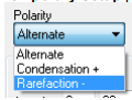
- Perform a baseline measure with the tube clamped. Make sure not to move the transducer when doing so.
- Select ear and intensity and start the measure. Clicks at the intensity level of 80-85dB nHL should be used.
- Monitor the EEG during testing to assure a collection with minimal noise.
- Monitor the response on the screen in the first few milliseconds – typically 2000 sweeps are conducted when measuring with the A/B enabled using an alternating stimuli – though the number of sweeps should not be used as stop criteria on its own
- If a cochlear microphonic is present, it is important to ensure that it is not a stimulus artifact. Make sure that the measure is reproducible and run a measure with the tube clamped.
Cochlear microphonic results
Below are two examples of cochlear microphonic responses. The upper is a case of ANSD, while the lower is measured on an individual with normal cochlear microphonic function. 
The upper example shows a cochlear microphonics of a baby with ANSD, showing rarefaction, condensation and base line with tube clamped (Stevens et al, 2011). The lower example shows a cochlear microphonic from an infant considered normal.
Patients with ANSD show an abnormal cochlear microphonic, seen as greater than normal amplitude of the response within the first milliseconds. In addition the latency of the cochlear microphonic duration is often longer than expected. Please note that the cochlear microphonic response itself is not sufficient documentation for ANSD and must be supported with an ABR recording to examine if the ABR response is present or absent.
Reporting
Choose the Report Icon ![]()
When complete, choose Save and Exit
References
Dallos P. (1983) Some electrical circuit properties of the organ of Corti. I. Analysis without reactive elements. Hear Res;12:89-120.
Stevens et al. (2011) Guidelines for Cochlear Microphonic Testing, NHS v. 2.0 Edited 2014.